Shoulder Tendon Anatomy : Fascias and spaces of the shoulder girdle: Anatomy | Kenhub : The common extensor tendon is a tendon that attaches to the lateral epicondyle of the.
Shoulder Tendon Anatomy : Fascias and spaces of the shoulder girdle: Anatomy | Kenhub : The common extensor tendon is a tendon that attaches to the lateral epicondyle of the.. The rotator cuff tendons attach to the deep rotator cuff muscles. Shoulder and pectoral region medicine 300 with mustafa/ulasli at gaziantep university. Shoulder radiology & anatomy at usuhs.mil. Notice that the supraspinatus tendon is parallel to the axis of the muscle. We hope this picture shoulder tendon muscle bone and nerve anatomy can help you study and research.
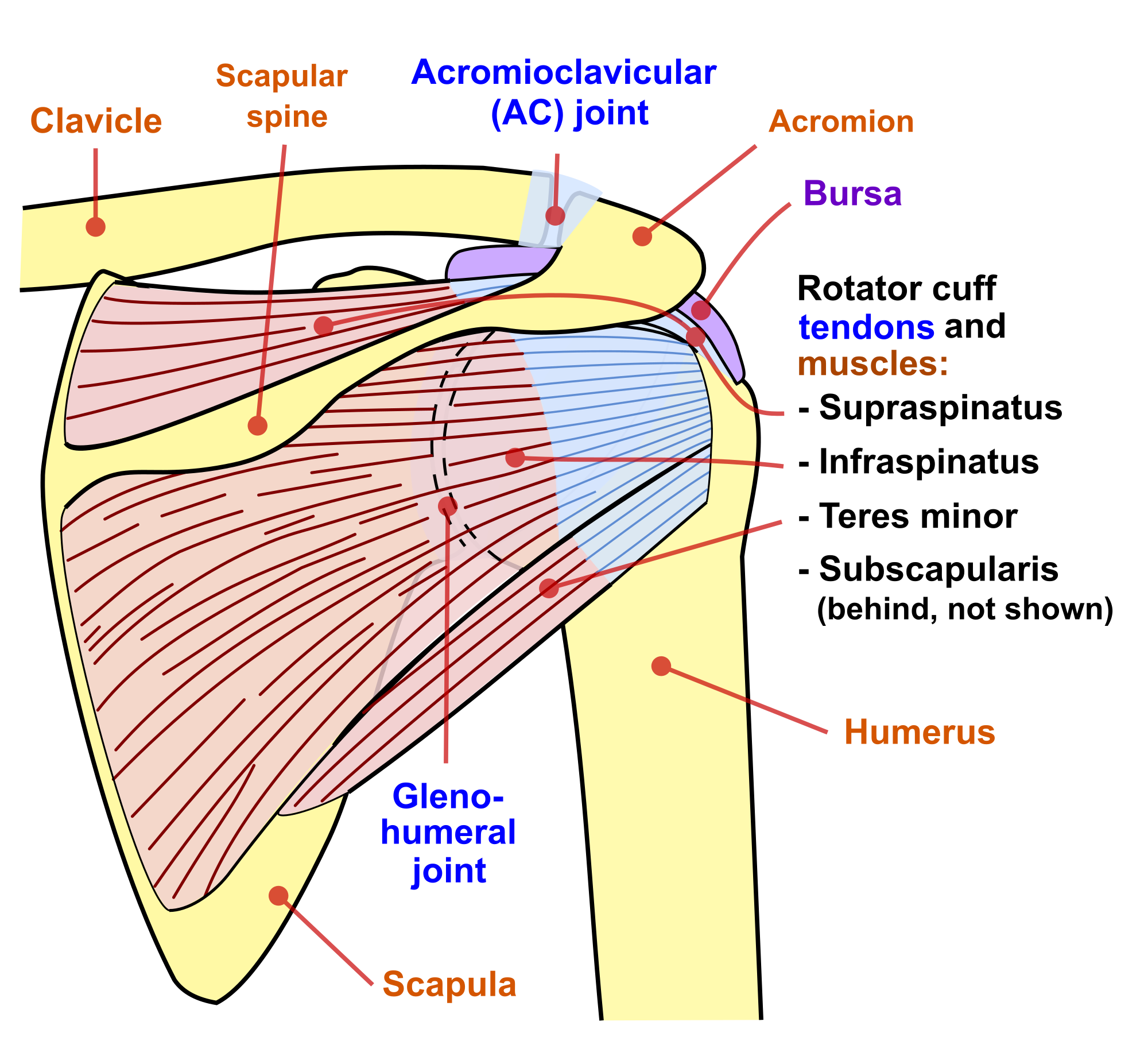
The bursa acts to cushion and reduce friction during motion between the overlying bone of the acromion. In the video below, dr. The ac joint is a diarthrodial and synovial joint. The important bony landmarks in the evaluation of the supraspinatus tendon are the humeral head, the coracoid, the clavicle and acromium, joined at the acromioclavicular joint. In addition to shoulder dislocations, other common injuries include rotator cuff tendon tears and broken bones including the humerus and collar bone.

Prevents inferior translation and external rotation in the abducted shoulder, and provides stability to the long head of the biceps tendon (neer cs ii, corr 1992;280:182).
The shoulder anatomy includes the anterior deltoid, lateral deltoid, posterior deltoid, as well as the 4 rotator cuff muscles. The subacromial bursa lies on the superior aspect of the supraspinatus tendon (see the images below). The multiple ligaments and tendons around the shoulder must be strong to bind the shoulder joints together and encapsulate them in a tough but flexible structure. Related online courses on physioplus. Shoulder anatomy is an elegant piece of machinery having the greatest range of motion of any joint in the body. The most common shoulder injuries involve the muscles, ligaments, cartilage, and tendons, rather than the bones. The shoulder is one of the largest and most complex joints in the body. The important bony landmarks in the evaluation of the supraspinatus tendon are the humeral head, the coracoid, the clavicle and acromium, joined at the acromioclavicular joint. Due to its complex anatomy the shoulder is prone to injuries and to degenerative wear and tear such as An image depicting shoulder anatomy can be seen below. Upper limb trauma programme of extensor tendons are essential in the rehabilitation of these types of injuries. It can help you understand our world more detailed and specific. Functional anatomy of the shoulder.
Shoulder anatomy is a remarkable combination of strong bones, flexible ligaments and tendons, and reinforcing cartilage and muscles. The subacromial bursa lies on the top portion of the supraspinatus tendon. In addition to shoulder dislocations, other common injuries include rotator cuff tendon tears and broken bones including the humerus and collar bone. Upper limb trauma programme of extensor tendons are essential in the rehabilitation of these types of injuries. The multiple ligaments and tendons around the shoulder must be strong to bind the shoulder joints together and encapsulate them in a tough but flexible structure.
Knowledge of the shoulder will help you understand the different shoulder problems.
The long head of biceps (lhb) is a very important tendon that travels through the shoulder joint (glenohumeral joint). All of these components of the shoulder, along with the muscles of the upper body, work together to manage the stress the shoulder receives as you extend, flex, lift, and throw. The bursa acts to cushion and reduce friction during motion between the overlying bone of the acromion. Related online courses on physioplus. Shoulder and pectoral region medicine 300 with mustafa/ulasli at gaziantep university. Look for an os acromiale. The long head biceps tendon travels through the shoulder joint making it more prone to injury such as a partial tear, rupture, or tendonitis. Notice that the supraspinatus tendon is parallel to the axis of the muscle. It can help you understand our world more detailed and specific. The shoulder joint (glenohumeral joint) is a ball and socket joint between the scapula and the humerus. Due to its complex anatomy the shoulder is prone to injuries and to degenerative wear and tear such as If symptoms cannot be relieved by. Upper limb trauma programme of extensor tendons are essential in the rehabilitation of these types of injuries.
The ac joint is a diarthrodial and synovial joint. The shoulder joint is formed where the humerus (upper arm bone) fits into the scapula. This is not always the case. Due to its complex anatomy the shoulder is prone to injuries and to degenerative wear and tear such as In addition to shoulder dislocations, other common injuries include rotator cuff tendon tears and broken bones including the humerus and collar bone.

Specifically, the four rotator cuff muscles include the following
The human shoulder is made up of three bones: The shoulder joint (glenohumeral joint) is a ball and socket joint between the scapula and the humerus. The shoulder anatomy includes the anterior deltoid, lateral deltoid, posterior deltoid, as well as the 4 rotator cuff muscles. Robin smithuis and henk jan van der woude. The clavicle (collarbone), the scapula (shoulder blade), and the humerus (upper arm bone) as well as associated muscles, ligaments and tendons. In addition to shoulder dislocations, other common injuries include rotator cuff tendon tears and broken bones including the humerus and collar bone. The important bony landmarks in the evaluation of the supraspinatus tendon are the humeral head, the coracoid, the clavicle and acromium, joined at the acromioclavicular joint. The long head of biceps (lhb) is a very important tendon that travels through the shoulder joint (glenohumeral joint). Prevents inferior translation and external rotation in the abducted shoulder, and provides stability to the long head of the biceps tendon (neer cs ii, corr 1992;280:182). Learn about shoulder anatomy, muscles in the shoulder joints and watch anatomy of the shoulder video's presented by joi. A bursa acts as a cushion between the tendons, muscles and bones, reducing friction and allowing smooth movement. Anatomy is the amazing science. This group of muscles lies just outside the shoulder joint.
Komentar
Posting Komentar